MRI
Jump to navigation
Jump to search
from chapter 1 of The Statistical Analysis of Functional MRI Data http://www.springerlink.com/content/978-0-387-78191-4/#section=212959&page=1&locus=0
Particles and Atoms
- atomic number - number of protons, determines kind of atom
- atomic weight - number of protons and neutrons, determines isotope
- spin
- 0 if atomic number and atomic weight are both even
- half-integer if atomic weight odd
- integer if atomic number odd, atomic weight even
1H (proton) has spin 1/2 and is the predominant subject of MRI
Fields and Precession
- one Tesla = 10,000 Gauss
- earth's magnetic field is 0.5 Gauss or 50 microTesla
- Larmor equation - atoms of nonzero spin absorb electromagnetic radiation of frequency <math>\omega</math>
- <math>\omega = \gamma B_0</math>
- <math>\gamma</math> is gyromagnetic ratio of the nucleus
- For a 3T magnet studying 1H nuclei, <math>\gamma</math> is 42.58 MHz/T, and <math>\omega</math> is 127.74 MHz (radio waves of length 2.347 m)
- particles with spin naturally precess around an axis aligned with the magnetic field
- Zeeman effect - as field strength goes up, proportionally more protons align parallel to field than anti-parallel
Acquiring Images
- A gradient coil induces a small gradient to the background field to give each position of the brain its own resonance frequency
- gradient field may cause peripheral nerve stimulation
- An RF pulse stimulates just those protons at exactly the right frequency to flip and precess in concert.
- RF pulse will heat tissue (SAR = specific absortion rate measured in Watts/kg)
- As the spins relax back to the low-energy parallel state, they emit radiation that gets picked up by receiver coils
- by applying a gradient that is uniform along one axis, parallel slices can be selected by incrementing the frequency
Anatomy of an MRI machine
- The subject lies within a cylindrical tube called the bore
- Immediately surrounding this is a cylinder called the body coil. It can send and receive RF pulses.
- Immediately surrounding the body coil is the gradient coil, which generates the x, y, and z gradient fields
- Around the gradient coil is the superconducting B0 coil, which generates the high magnetic field. The vast majority of the MRI's expense lies within this superconducting solenoid, which must be chilled with liquid helium to around 4 Kelvins.
Relaxation Times
- T1 spin-lattice or longitudinal relaxation time - time required by the z component of the tissue magnetization to return to 63% of its original value
- <math>M_z = M_0 (1 - e^{ - t/T_1} ).</math>
- T2 spin-spin or transverse relaxation time - time required by the transverse component of the magnetization to return to 37% of its original value
- <math>M_{xy} = M_0 e^{ - t/T_2}</math>
- <math>T_2^*</math> free induction decay (FID) relaxation time - decay constant for an exponentially damped sinusoidal
- In general, <math>T_2^* < T_2 < T_1</math>
| values at 1.5T (ms) | T1 | T2 |
|---|---|---|
| gray matter | 900 | 100 |
| white matter | 600 | 80 |
| cerebrospinal fluid | 4000 | 2000 |
fMRI
(see also fMRI)
- Measures increase in oxygenated blood flow to regions with active neurons
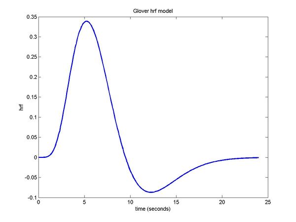
- hemodynamic response function (HRF) - the ratio of deoxygenated to oxygenated blood as a function of time
- blood oxygenation level dependent (BOLD) effect - deoxygenated blood has a magnetic susceptibility that is 20% greater than that of oxygenated blood
- the density of hydrogen nuclei at a given point is the inverse Fourier transform of the measured magnetizations over specific frequencies and phases
- the signal from the center of k-space overwhelms everything else and has to be damped down to see the rest
- repetition time (TR, eg 2s) is the time between consecutive scans of the brain (in which a full set of slices is obtained)
- TR may be varied to optimize contrast between a particular pair of tissue types
- echo time (TE) is the time between the RF pulse and the peak of the echoing response signal (how is this under the control of the experimenter?)
- T1 - weighted images (intermediate TR, short TE) emphasize white and gray matter
- T2 - weighted images (long TR, intermediate TE) emphasize cerebrospinal fluid, good for structurals
- <math>T_2^*</math> - weighted images (long TR, intermediate TE) emphasize deoxygenated blood
- signal to noise ratio (SNR) is proportional to the volume of a voxel and <math>\sqrt{\frac{N_y NEX}{BW}}</math>, where
- <math>N_y</math> is the number of phase encoding steps (i.e. number of voxels in the y direction)
- NEX is the number of excitations (number of times the scan is repeated)
- BW is the bandwidth (?)
